Our Services

We offer a full range of
Ultrasound, CT and MRI Scans.

Imaging Techniques

Ultrasound (US)

Ultrasound (US)

At Radiology Insights, we use US to help identify the location of your symptoms and assess the problem you are experiencing as well as using US guidance for any injections we do to optimise the delivery of therapeutic medications to a specific target.

Your scan will be performed by a highly experienced Consultant Radiologists using the state-of-the-art Canon Aplio Prism i-700 Ultrasound machine.

Ultrasound technology uses high-frequency sound waves to produce real-time images of soft tissue without radiation. It is safe and commonly used in antenatal and neonatal imaging.

Abdominal Ultrasound Scan

An abdominal Ultrasound scan is a painless and non-invasive procedure that uses sound waves to create images of the organs and structures in your abdomen.

One of the main benefits of an abdominal ultrasound scan is that it helps doctors evaluate the health and condition of your abdominal organs, such as the liver, gallbladder, kidneys, pancreas, and spleen. The scan can help identify abnormalities, such as tumours, cysts, or stones, and provide valuable information for diagnosing conditions like liver disease, gallstones, kidney stones, or pancreatitis.

The procedure is also valuable in examining blood flow to the abdominal organs. It can help detect any issues with blood vessels or identify signs of reduced blood flow, which can indicate problems such as blood clots or narrowing of blood vessels.

Is there is any radiation exposure?

An ultrasound of the testes and scrotum is a non-invasive and painless procedure that can provide valuable insights without radiation exposure.

Is it safe?

an ultrasound of the testes and scrotum is a safe procedure. It is non-invasive, painless, and does not involve any radiation.

Why might I need this type of scan?

Indication for abdminal ultrasound scan may include:

Abdominal Pain

The scan can help identify the source of unexplained pain of discomfort in the abdomen.

Liver or gallbladder problems

The scan can evaluate the liver for conditions such as fatty liver disease, hepatitis, or cirrhosis. It can also detect gallstones or check the gallbladder for inflammation or infection.

Digestive issues

It can be used to investigate issues like unexplained weight loss, bloating, or changes in bowel movements.

Kidney or bladder problems

It helps in assessing the kidneys for abnormalities, such as stones systsm or infections. The scan can also evaluate the bladder for issues like urinary tract infections or obstructions.

Pancreatic or spleen issues

It can provide valuable information about the pancreas or spleen, helping to detect conditions like pancreatitis or enlarged spleen.

Abdominal Ultrasound Scan

An Ultrasound of the testes and scrotum is a non-invasive imaging test that uses sound waves to create detailed images of the structures within the male reproductive system.

One of the main benefits of an Ultrasound of the testes and scrotum is that it provides a clear view of the testicles, epididymis, and surrounding structures. The scan can help identify any abnormalities, such as tumours, cysts, inflammation, or fluid accumulation, providing valuable information for diagnosing conditions like testicular torsion, epididymitis, varicoceles, hydroceles, or testicular cancer.

The procedure is also useful in evaluating blood flow to the testicles. It can help detect any issues with blood vessels or identify signs of reduced blood flow, which can indicate problems such as testicular torsion or varicoceles.

Is there is any radiation exposure?

An Ultrasound scan is safe and does not involve exposure to ionising radiation.

Is it safe?

An Ultrasound is a safe procedure. It is non-invasive, painless and does not involve any radiation.

Why might I need this type of injection?

Indications for an Ultrasound of the testes and scrotum may include:

Testicular pain or discomfort

The scan can help identify the source of unexplained pain or discomfort in the testes or scrotum

Swelling or enlargement of the scrotum

It can be used to investigate scrotal swelling or enlargement and identify potential causes, such as hydroceles or hernias.

Evaluation of testicular masses or lumps

The scan can assess the characteristics and location of testicular masses, which can help diagnose conditions like testicular cancer or benign tumours.

Fertility concerns

It can help evaluate the structure and function of the testicles and assess potential causes of infertility, such as varicoceles.

Follow-up after testicular surgery or injury

An Ultrasound can be used to monitor the healing process and assess the success of surgical repairs or other interventions.

Ultrasound scan of the Kidneys and Urinary system

An Ultrasound of the kidney and urinary system is a non-invasive imaging test that uses sound waves to create detailed images of the structures within these organs.

One of the main benefits of an Ultrasound of the kidney and urinary system is that it provides a clear view of these structures, including the kidneys, ureters, bladder, and sometimes the prostate or uterus. The scan can help identify any abnormalities, such as kidney stones, cysts, tumours, or signs of infection, providing valuable information for diagnosing conditions like urinary tract infections, kidney disease, kidney stones, or urinary tract obstructions.

The procedure is also useful in evaluating blood flow to the kidneys. It can help detect any issues with blood vessels or identify signs of reduced blood flow, which can indicate problems such as renal artery stenosis or renal vein thrombosis.

Furthermore, an Ultrasound of the kidney and urinary system is a safe and painless procedure that does not involve radiation exposure, making it suitable for people of all ages, including pregnant women and children.

Is there is any radiation exposure?

An Ultrasound scan is safe and does not involve exposure to ionising radiation.

Is it safe?

An Ultrasound is a safe procedure. It is non-invasive, painless and does not involve any radiation.

Why might I need this type of injection?

Indications for an Ultrasound of the kidney and urinary system may include:

Kidney or urinary symptoms

The scan can help identify the source of unexplained symptoms such as flank pain, blood in the urine, frequent urination, or difficulty urinating.

Urinary tract infections

It can be used to investigate recurrent or persistent urinary tract infections and identify any structural abnormalities contributing to the condition.

Kidney stones

The scan can help identify the presence, size, and location of kidney stones, aiding in their diagnosis and treatment planning.

Monitoring kidney function

It can be used to assess the size and structure of the kidneys and monitor changes in kidney function over time.

Evaluation of urinary tract obstructions

The scan can help identify any blockages or obstructions in the urinary tract, which can cause problems with urine flow.

Ultrasound of the Neck/Thyroid

An Ultrasound of the neck and thyroid is a non-invasive imaging test that uses sound waves to create detailed images of the structures within the neck, including the thyroid gland.

One of the main benefits of an Ultrasound of the neck and thyroid is that it provides a clear view of the structures in the neck region, including the thyroid gland, lymph nodes, and surrounding tissues. The scan can help identify any abnormalities, such as nodules, cysts, or enlarged lymph nodes, providing valuable information for diagnosing conditions like thyroid nodules, thyroiditis, or lymphadenopathy.

The procedure is also useful in evaluating the size, shape, and characteristics of the thyroid gland itself. It can help detect any irregularities in the glands structure, such as nodules or signs of inflammation.

An Ultrasound of the neck and thyroid is a safe and painless procedure that does not involve radiation exposure, making it suitable for people of all ages.

Is there is any radiation exposure?

An Ultrasound scan is safe and does not involve exposure to ionising radiation.

Is it safe?

An Ultrasound is a safe procedure. It is non-invasive, painless and does not involve any radiation.

Why might I need this type of injection?

Indications for an Ultrasound of the kidney and urinary system may include:

Thyroid nodules or goitre

The scan can help assess the size, number, and characteristics of thyroid nodules or goitre, aiding in their diagnosis and determining if further evaluation or treatment is needed

Thyroid function monitoring:

It can be used to assess the overall health and function of the thyroid gland, especially in cases of thyroid disorders such as hypothyroidism or hyperthyroidism.

Thyroid cancer evaluation

The scan can help identify any suspicious nodules or abnormalities that may require further investigation to rule out or diagnose thyroid cancer.

Enlarged lymph nodes

It can help evaluate the size and characteristics of enlarged lymph nodes in the neck, which may indicate infection, inflammation, or other underlying conditions.

Ultrasound Shoulder

An Ultrasound of the shoulder is a safe and non-invasive imaging test that uses sound waves to create detailed images of the structures within your shoulder joint.


One of the main benefits of an Ultrasound of the shoulder is that it provides a clear view of the soft tissues in and around the shoulder joint. This includes the muscles, tendons, ligaments, and bursae. The scan can help identify any abnormalities, such as tears, inflammation, or fluid accumulation, providing valuable information for diagnosing conditions like rotator cuff tears, tendonitis, bursitis, or shoulder impingement.

The procedure is also valuable in evaluating blood flow to the shoulder area. It can help detect any issues with blood vessels or identify signs of reduced blood flow, which can indicate problems such as blood clots or vascular abnormalities.

An Ultrasound of the shoulder is a dynamic imaging technique, meaning that the scan can be performed while you actively move your shoulder. This allows for real-time evaluation of the structures and can help assess any impingement or instability issues that may not be visible in static images alone.

Is there is any radiation exposure?

An Ultrasound scan is safe and does not involve exposure to ionising radiation.

Is it safe?

An Ultrasound is a safe procedure. It is non-invasive, painless and does not involve any radiation.

Why might I need this type of injection?

Indications for an Ultrasound of the shoulder may include:

Shoulder pain or discomfort

The scan can help identify the source of unexplained pain or discomfort in the shoulder area.

Suspected tendon or ligament tears

The scan can assess the integrity of the tendons and ligaments within the shoulder joint, helping to diagnose conditions such as rotator cuff tears or ligament injuries.

Follow-up after shoulder surgery or injury

An Ultrasound can be used to monitor the healing process and assess the success of surgical repairs or other interventions.

Limited range of motion

It can be used to investigate issues with shoulder mobility and identify any structural abnormalities contributing to restricted movement.

Evaluation of bursae

It can help assess the bursae, which are fluid-filled sacs that provide cushioning around the joint. The scan can identify any inflammation or fluid accumulation in the bursae, indicating conditions like bursitis.

Ultrasound Shoulder

An Ultrasound of the elbow is a non-invasive imaging test that uses sound waves to create detailed images of the structures within your elbow joint.


One of the main benefits of an Ultrasound of the elbow is that it provides a clear view of the soft tissues in and around the joint. This includes the muscles, tendons, ligaments, and bursae. The scan can help identify any abnormalities, such as tears, inflammation, or fluid accumulation, providing valuable information for diagnosing conditions like tendonitis, bursitis, or ligament injuries.

The procedure is also useful in evaluating blood flow to the elbow area. It can help detect any issues with blood vessels or identify signs of reduced blood flow, which can indicate problems such as blood clots or vascular abnormalities.

An Ultrasound of the elbow allows for dynamic imaging, meaning that the scan can be performed while you actively move your elbow. This real-time evaluation can help assess any impingement or instability issues and provide insights into the joint s overall function.

Is there is any radiation exposure?

An Ultrasound scan is safe and does not involve exposure to ionising radiation.

Is it safe?

An Ultrasound is a safe procedure. It is non-invasive, painless and does not involve any radiation.

Why might I need this type of injection?

Indications for an Ultrasound of the elbow may include:

Elbow pain or discomfort

The scan can help identify the source of unexplained pain or discomfort in the elbow area.

Tendon or ligament injuries

The scan can assess the integrity of the tendons and ligaments within the elbow joint, helping to diagnose conditions such as tennis elbow (lateral epicondylitis), golfer s elbow (medial epicondylitis), or ligament tears.

Evaluation of nerve entrapment:

The scan can provide insights into nerve-related issues, such as cubital tunnel syndrome or radial tunnel syndrome.

Limited range of motion

It can be used to investigate issues with shoulder mobility and identify any structural abnormalities contributing to restricted movement.

Bursitis

It can help assess the bursae around the elbow joint and identify any inflammation or fluid accumulation, indicating conditions like olecranon bursitis.

Ultrasound Wrist

An Ultrasound of the wrist is a non-invasive imaging test that uses sound waves to create detailed images of the structures within your wrist joint.


One of the main benefits of an Ultrasound of the wrist is that it provides a clear view of the soft tissues in and around the joint. This includes the tendons, ligaments, muscles, and other structures. The scan can help identify any abnormalities, such as tears, inflammation, or fluid accumulation, providing valuable information for diagnosing conditions like tendonitis, ligament injuries, ganglion cysts, or arthritis.

The procedure is also useful in evaluating blood flow to the wrist area. It can help detect any issues with blood vessels or identify signs of reduced blood flow, which can indicate problems such as blood clots or vascular abnormalities.

An Ultrasound of the wrist allows for dynamic imaging, meaning that the scan can be performed while you actively move your wrist. This real-time evaluation can help assess any impingement, instability, or joint alignment issues and provide insights into the joint s overall function.

Is there is any radiation exposure?

An Ultrasound scan is safe and does not involve exposure to ionising radiation.

Is it safe?

An Ultrasound is a safe procedure. It is non-invasive, painless and does not involve any radiation.

Why might I need this type of injection?

Indications for an Ultrasound of the wrist may include:

Wrist pain or discomfort

The scan can help identify the source of unexplained pain or discomfort in the wrist area.

Tendon or ligament injuries

The scan can assess the integrity of the tendons and ligaments within the wrist joint, helping to diagnose conditions such as wrist tendonitis, ligament tears, or sprains.

Arthritis

The scan can provide insights into the presence and severity of arthritis in the wrist joint.

Limited range of motion

It can be used to investigate issues with wrist mobility and identify any structural abnormalities contributing to restricted movement.

Evaluation of ganglion cysts

It can help assess the size, location, and characteristics of ganglion cysts, which are fluid-filled lumps that can develop near joints.

Ultrasound Hand

An Ultrasound of the hand is a non-invasive imaging test that uses sound waves to create detailed images of the structures within your hand.


One of the main benefits of an Ultrasound of the hand is that it provides a clear view of the soft tissues in and around the hand, including tendons, ligaments, muscles, and other structures. The scan can help identify any abnormalities, such as tears, inflammation, or fluid accumulation, providing valuable information for diagnosing conditions like tendonitis, ligament injuries, cysts, or arthritis.

An Ultrasound of the hand allows for dynamic imaging, meaning that the scan can be performed while you actively move your hand. This real-time evaluation can help assess any impingement, instability, or joint alignment issues and provide insights into the hand’s overall function.

Is there is any radiation exposure?

An Ultrasound scan is safe and does not involve exposure to ionising radiation.

Is it safe?

An Ultrasound is a safe procedure. It is non-invasive, painless and does not involve any radiation.

Why might I need this type of injection?

Indications for an Ultrasound of the hand may include:

Hand pain or discomfort

The scan can help identify the source of unexplained pain or discomfort in the hand.

Tendon or ligament injuries

The scan can assess the integrity of the tendons and ligaments within the hand, helping to diagnose conditions such as trigger finger, De Quervains tenosynovitis, ligament tears, or sprains.

Arthritis

The scan can provide insights into the presence and severity of arthritis in the hand joints.

Limited range of motion

It can be used to investigate issues with hand mobility and identify any structural abnormalities contributing to restricted movement.

Evaluation of cysts or masses

It can help assess the characteristics and location of cysts, ganglion cysts, or other masses within the hand.

Ultrasound Hip

An Ultrasound of the hip is a non-invasive imaging test that uses sound waves to create detailed images of the structures within your hip joint.


One of the main benefits of an Ultrasound of the hip is that it provides a clear view of the soft tissues in and around the joint, including the muscles, tendons, ligaments, and other structures. The scan can help identify any abnormalities, such as tears, inflammation, or fluid accumulation, providing valuable information for diagnosing conditions like hip tendonitis, bursitis, labral tears, or snapping hip syndrome.

The procedure is also useful in evaluating blood flow to the hip area. It can help detect any issues with blood vessels or identify signs of reduced blood flow, which can indicate problems such as vascular abnormalities.

Furthermore, an Ultrasound of the hip allows for dynamic imaging, meaning that the scan can be performed while you actively move your hip joint. This real-time evaluation can help assess any impingement, instability, or joint alignment issues and provide insights into the hip s overall function.

Is there is any radiation exposure?

An Ultrasound scan is safe and does not involve exposure to ionising radiation.

Is it safe?

An Ultrasound is a safe procedure. It is non-invasive, painless and does not involve any radiation.

Why might I need this type of injection?

Indications for an Ultrasound of the hip may include:

Hip pain or discomfort

The scan can help identify the source of unexplained pain or discomfort in the hip joint.

Tendon or ligament injuries

The scan can assess the integrity of the tendons and ligaments within the hip joint, helping to diagnose conditions such as hip tendonitis, ligament tears, or strains.

Labral tears

The scan can provide insights into the labrum, which is a cartilage structure within the hip joint. The scan can help identify labral tears, which are common hip injuries.

Limited range of motion

It can be used to investigate issues with hip mobility and identify any structural abnormalities contributing to restricted movement.

Evaluation of bursae

It can help assess the bursae around the hip joint and identify any inflammation or fluid accumulation, indicating conditions like trochanteric bursitis.

Ultrasound Knee

An Ultrasound of the knee is a non-invasive imaging test that uses sound waves to create detailed images of the structures within your knee joint.


One of the main benefits of an Ultrasound of the knee is that it provides a clear view of the soft tissues in and around the joint. This includes the tendons, ligaments, muscles, and other structures. The scan can help identify any abnormalities, such as tears, inflammation, or fluid accumulation, providing valuable information for diagnosing conditions like tendonitis, ligament injuries, bursitis, or meniscus tears.

The procedure is also useful in evaluating blood flow to the knee area. It can help detect any issues with blood vessels or identify signs of reduced blood flow, which can indicate problems such as blood clots or vascular abnormalities.

An Ultrasound of the knee allows for dynamic imaging, meaning that the scan can be performed while you actively move your knee joint. This real-time evaluation can help assess any impingement, instability, or joint alignment issues and provide insights into the knee s overall function.

Is there is any radiation exposure?

An Ultrasound scan is safe and does not involve exposure to ionising radiation.

Is it safe?

An Ultrasound is a safe procedure. It is non-invasive, painless and does not involve any radiation.

Why might I need this type of injection?

Indications for an Ultrasound of the knee may include:

Knee pain or discomfort

The scan can help identify the source of unexplained pain or discomfort in the knee joint.

Evaluation of tendon or ligament injuries

The scan can assess the integrity of the tendons and ligaments within the knee joint, helping to diagnose conditions such as patellar tendonitis, ACL tears, or sprains.

Follow-up after knee surgery or injury

An Ultrasound can be used to monitor the healing process and assess the success of surgical repairs or other interventions.

Swelling or inflammation

It can be used to investigate knee swelling or inflammation and identify potential causes, such as bursitis or synovitis.

Meniscus tears

It can provide insights into the meniscus, which is a cartilage structure within the knee joint. The scan can help identify meniscus tears, which are common knee injuries.

Ultrasound Foot

An Ultrasound of the foot is a non-invasive imaging test that uses sound waves to create detailed images of the structures within your foot.


One of the main benefits of an Ultrasound of the foot is that it provides a clear view of the soft tissues in and around the foot, including tendons, ligaments, muscles, and other structures. The scan can help identify any abnormalities, such as tears, inflammation, or fluid accumulation, providing valuable information for diagnosing conditions like plantar fasciitis, Achilles tendonitis, ligament injuries, or Morton’s neuroma.

The procedure is also useful in evaluating blood flow to the foot. It can help detect any issues with blood vessels or identify signs of reduced blood flow, which can indicate problems such as peripheral artery disease or vascular abnormalities.

Furthermore, an Ultrasound of the foot allows for dynamic imaging, meaning that the scan can be performed while you actively move your foot. This real-time evaluation can help assess any impingement, instability, or joint alignment issues and provide insights into the foot’s overall function.

Is there is any radiation exposure?

An Ultrasound scan is safe and does not involve exposure to ionising radiation.

Is it safe?

An Ultrasound is a safe procedure. It is non-invasive, painless and does not involve any radiation.

Why might I need this type of injection?

Indications for an Ultrasound of the foot may include:

Foot pain or discomfort

The scan can help identify the source of unexplained pain or discomfort in the foot.

Evaluation of tendon or ligament injuries

The scan can assess the integrity of the tendons and ligaments within the foot, helping to diagnose conditions such as plantar fasciitis, Achilles tendonitis, or ligament sprains.

Evaluation of masses or cysts

The scan can help assess the characteristics and location of masses or cysts within the foot.

Swelling or inflammation

It can be used to investigate foot swelling or inflammation and identify potential causes, such as bursitis or tendonitis.

Nerve-related issues

It can provide insights into nerve-related problems such as Morton s neuroma or tarsal tunnel syndrome.

Dynamic Ultrasound of the Thorax and Ribs (DUTR)

Dynamic Thoracic Ultrasound takes advantage of real-time US imaging to allow “dynamic” assessment of the chest wall during specific manoeuvres. This is particularly advantageous in the imaging of ribs and the chest wall as certain pathologies such as slipping rib can only be triggered with specific movements.

At RI London we have refined and improved our techniques in Dynamic Ultrasounds and developed our own techniques and methodologies which have resulted in greater diagnostic accuracy and reproducibility. This has been particularly effective in the assessment and diagnosis of Slipping Rib Syndrome.

At RI London, we assess rib and chest wall pain in a “whole system” approach. This means that we understand symptoms to be a result of a complex dysfunction of thoracic, lower back and abdominal wall stability (“Trunk stability”).

Like all imaging techniques, US and DTUS do have limitations. The technique is user-dependent – that is to say, the diagnostic accuracy and reproducibility are highly dependent on the skill of the operator.

Ultrasound Guided Injections

An Ultrasound-guided injection is a minimally invasive medical procedure that uses Ultrasound imaging to guide the injection of medication directly into a specific area of the body. This technique is used to provide relief for a variety of medical conditions, including joint pain, muscle pain, and inflammation.

During an Ultrasound-guided injection, one of our Consultant Radiologists uses Ultrasound imaging to precisely locate the area of the body that needs treatment. This allows them to see the exact location of the injection site and ensure that the medication is delivered to the correct area.

Ultrasound guided injections

An Ultrasound-guided joint injection is a procedure in which medication, such as a corticosteroid or a local anaesthetic, is injected directly into a joint under the guidance of an Ultrasound machine. The Ultrasound allows the Consultant to visualise the joint and surrounding structures in real-time, ensuring accurate needle placement.


An Ultrasound-guided joint injection has several advantages over a blind or unguided injection. The use of Ultrasound allows for real-time visualisation of the joint, ensuring accurate needle placement and reducing the risk of complications. It also allows for better targeting of specific areas within the joint, such as inflamed bursae or small joint spaces.


Is there is any radiation exposure?

An Ultrasound scan is safe and does not involve exposure to ionising radiation.

Is it safe?

An Ultrasound is a safe procedure. It is non-invasive, painless and does not involve any radiation.

Benefits of Ultrasound-Guided Injections

Increased Accuracy

The real-time imaging provided by Ultrasound ensures that the injection is precisely delivered to the targeted area, enhancing the effectiveness of the treatment.

Minimally Invasive

Ultrasound-guided injections are minimally invasive, meaning there is no need for surgical incisions, leading to faster recovery times and minimal risk of complications.

Safety

Ultrasound guidance reduces the risk of complications by avoiding structures such as nerves, blood vessels, and other sensitive areas.

Reduced Pain and Discomfort

Because the procedure is more accurate, patients often experience less pain and fewer side effects compared to traditional blind injections.

Versatility

This technique can be used to treat a variety of conditions affecting different parts of the body, including the shoulders, knees, hips, and spine.

What to Expect During the Procedure?

Preparation

The area to be injected is cleansed, and a local anaesthetic may be applied to minimise discomfort.

Injection

Under Ultrasound guidance, the physician carefully inserts the needle and administers the medication directly into the targeted area.

Imaging

An Ultrasound probe is used to identify the precise location for the injection.

Post-Procedure

After the injection, the patient may be monitored briefly and given instructions for aftercare. Most people can resume normal activities shortly after the procedure.

Aftercare and Recovery

After an Ultrasound-guided injection, patients may experience temporary soreness or mild discomfort at the injection site. It is generally recommended to avoid strenuous activity for a few days following the procedure. Applying ice and taking over-the-counter pain relievers can help manage any post-procedural pain. Most patients experience relief within a few days, although the full effects of the injection may take a week or more to become evident.

Is Ultrasound-Guided Injection Right for You

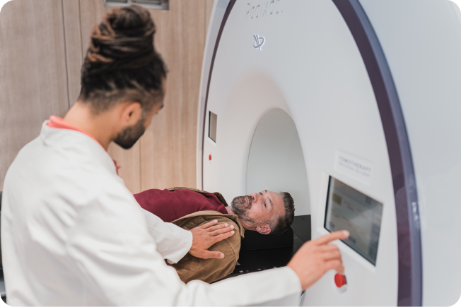
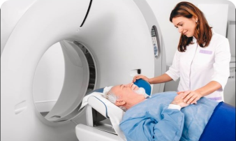
If you are experiencing chronic pain or inflammation that has not responded to conservative treatments such as physical therapy, rest, or medications, an Ultrasound-guided injection may be an effective option for you. It is important to consult with a healthcare professional to determine if this procedure is appropriate for your specific condition and medical history.

Nerve Block

A nerve block is a medical procedure that involves the injection of an anaesthetic or anti-inflammatory medication directly into a specific nerve or group of nerves to relieve pain. This procedure can be used for both diagnostic and therapeutic purposes, making it a versatile tool in pain management. Nerve blocks are commonly used to manage acute or chronic pain conditions, including pain associated with surgery, injury, or chronic medical conditions like arthritis or neuropathy.


A nerve block works by interrupting pain signals traveling along a nerve pathway to the brain. The injection of anaesthetic temporarily “blocks” these signals, which provides immediate relief from pain. In some cases, additional medications, such as steroids, may be injected to reduce inflammation and provide longer-lasting pain relief.


Is there is any radiation exposure?

An Ultrasound scan is safe and does not involve exposure to ionising radiation.

Is it safe?

An Ultrasound is a safe procedure. It is non-invasive, painless and does not involve any radiation.

How Does a Nerve Block Work?

A nerve block works by interrupting pain signals traveling along a nerve pathway to the brain. The injection of anaesthetic temporarily “blocks” these signals, which provides immediate relief from pain. In some cases, additional medications, such as steroids, may be injected to reduce inflammation and provide longer-lasting pain relief.

Types of Nerve Blocks

Intercostal Nerve Block

Intercostal nerves are located between the ribs and are responsible for transmitting pain signals from the chest and upper abdominal region to the brain. During an intercostal nerve block, a local anaesthetic is injected directly into the area around these nerves. The anaesthetic temporarily 'blocks' the transmission of pain signals, providing significant pain relief

Sympathetic Nerve Block

This block targets the sympathetic nervous system and is used to treat pain conditions such as complex regional pain syndrome (CRPS).

Facet Joint Block

This block targets the small joints located between each set of vertebrae in the spine, providing relief from neck or back pain.

Arthritis

Pain relief for inflammatory conditions affecting joints, such as rheumatoid arthritis or osteoarthritis.

Peripheral Nerve Block

This type targets specific nerves outside the spinal cord, such as those in the arms, legs, or face. It is often used for surgeries on the extremities.

Epidural and Spinal Nerve Blocks

These blocks are injected near the spinal cord and are commonly used for pain relief during labour and delivery or for surgeries involving the lower back and lower extremities.

Trigger Point Injections

These injections target specific 'trigger points' within muscles that cause pain, often used to treat myofascial pain syndrome.

Headaches and Migraines

Targeted nerve blocks can provide relief from severe headaches or migraines that do not respond to other treatments.

Benefits of Nerve Blocks

Targeted Pain Relief

Nerve blocks provide localized pain relief, making them highly effective for treating specific areas of pain.

Rapid Pain Relief

Many patients experience immediate pain relief following the injection, which can last for several days to several months, depending on the type of block and medication used.

Versatility

Nerve blocks can be used for both diagnostic purposes (to determine the source of pain) and therapeutic purposes (to provide long-term pain relief).

Minimally Invasive

The procedure is less invasive than surgery and can often be performed on an outpatient basis, allowing patients to go home the same day.

Reduced Need for Opioids

By providing effective pain relief, nerve blocks can reduce the need for opioid pain medications, decreasing the risk of dependency and side effects.

What to Expect During the Procedure?

Injection

Many patients experience immediate pain relief following the injection, which can last for several days to several months, depending on the type of block and medication used.

Versatility

The physician carefully inserts the needle and injects the anaesthetic or other medications around the targeted nerve.

Imaging Guidance

In many cases, imaging techniques such as Ultrasound or fluoroscopy are used to guide the needle to the precise location of the targeted nerve or nerve group.

Post-Procedure

After the injection, the patient may be monitored briefly and given instructions for aftercare. Most people can resume normal activities shortly after the procedure.

Aftercare and Recovery Is a Nerve Block Right for You?

Nerve blocks can be an effective option for individuals suffering from chronic or acute pain that has not responded well to other treatments. If you are experiencing persistent pain that affects your quality of life, a nerve block may provide the relief you need. It is essential to consult with a healthcare provider to determine if this procedure is appropriate for your specific condition and medical history.

Magnetic Resonance Imaging

Magnetic Resonance Imaging (MRI) is a sophisticated and technologically advanced technique that utilises powerful magnets to produce high-resolution images without radiation. It offers superior tissue visualisation, detailed structural and functional characterisation, and continuous advancements in technology and applications

At RI, we utilise cutting-edge 3T scanners with AI technology for the highest quality images. Our detailed scans use small segment slice images, unlike cheaper scans that use lower quality scanners and larger segment slices, potentially losing critical diagnostic information.

MRI Brain

An MRI (Magnetic Resonance Imaging) of the brain is a non-invasive diagnostic test that uses a powerful magnetic field and radio waves to create detailed images of the structures within your brain.


One of the main benefits of an MRI of the brain is that it provides a highly detailed view of the brain s anatomy. The scan can help identify any abnormalities, such as tumours, lesions, or signs of stroke, providing valuable information for diagnosing conditions like brain tumours, multiple sclerosis, stroke, or traumatic brain injury.

The procedure is also useful in evaluating blood flow to the brain. It can help detect any issues with blood vessels or identify signs of reduced blood flow, which can indicate problems such as blocked arteries or aneurysms.

Furthermore, an MRI of the brain provides a comprehensive evaluation of brain structures, including the cortex, white matter, ventricles, and other regions. It can help visualise the size, shape, and location of different brain structures, aiding in the diagnosis and treatment planning for various neurological conditions.

Is there is any radiation exposure?

An ultrasound of the testes and scrotum is a non-invasive and painless procedure that can provide valuable insights without radiation exposure.

Is it safe?

an ultrasound of the testes and scrotum is a safe procedure. It is non-invasive, painless, and does not involve any radiation.

Why might I need this type of scan?

Indications for an MRI of the brain may include:

Headaches or migraines

The scan can help identify the cause of recurrent headaches or migraines and rule out underlying structural abnormalities.

Neurological disorders

The scan can provide insights into conditions such as multiple sclerosis, brain tumours, Parkinson s disease, Alzheimer s disease, or stroke.

Follow-up after brain surgery or treatment

An MRI can be used to monitor the effectiveness of treatments, such as radiation therapy or surgery, and evaluate the progress of the healing process.

Seizures or epilepsy

It can be used to investigate the source of seizures and identify any brain abnormalities contributing to the condition.

Traumatic brain injury

It can help assess the extent of brain injury following a traumatic event, such as a concussion or head trauma.

MRI Joints

MRI (Magnetic Resonance Imaging) can be used to perform scans of joints in the body. MRI of joints is a non-invasive imaging technique that uses a powerful magnetic field and radio waves to create detailed images of the internal structures of the joint.


The MRI scan of a joint can provide valuable information about the bones, cartilage, tendons, ligaments, and surrounding soft tissues , aiding in diagnosis, treatment planning, and monitoring of joint conditions.

Is there is any radiation exposure?

An ultrasound of the testes and scrotum is a non-invasive and painless procedure that can provide valuable insights without radiation exposure.

Is it safe?

an ultrasound of the testes and scrotum is a safe procedure. It is non-invasive, painless, and does not involve any radiation.

Why might I need this type of scan?

It can help diagnose various conditions and injuries affecting the joint, including:

Joint pain or inflammation

An MRI can help identify the cause of unexplained joint pain or inflammation, such as arthritis, bursitis, or synovitis.

Cartilage damage

The scan can detect cartilage abnormalities, such as cartilage tears or degeneration, which are common in conditions like osteoarthritis or meniscus tears.

Evaluation of joint replacements

MRI can be used to assess the condition of joint replacements, such as artificial hips or knees, to identify any issues such as loosening or infection.

Ligament or tendon injuries

It can assess the integrity of ligaments and tendons within the joint, helping to diagnose conditions like ligament tears (such as ACL or MCL tears) or tendonitis.

Joint infections or abscesses

MRI can help detect the presence of infections or abscesses within the joint.

MRI spine

MRI (Magnetic Resonance Imaging) of the spine is a non-invasive imaging technique that uses a powerful magnetic field and radio waves to create detailed images of the structures within the spine. It is commonly used to evaluate various conditions and injuries affecting the spinal column.


MRI of the spine can provide valuable information about the bones, discs, spinal cord, nerve roots, and surrounding soft tissues.

Is there is any radiation exposure?

An ultrasound of the testes and scrotum is a non-invasive and painless procedure that can provide valuable insights without radiation exposure.

Is it safe?

an ultrasound of the testes and scrotum is a safe procedure. It is non-invasive, painless, and does not involve any radiation.

Why might I need this type of scan?

It can help diagnose and assess conditions such as:

Disc herniation

An MRI can reveal the location and severity of disc herniation, which occurs when the soft cushioning discs between the vertebrae bulge or rupture.

Degenerative disc disease

The scan can show the condition and health of the spinal discs, which may degenerate over time and contribute to pain and dysfunction.

Spinal cord or nerve compression

It can help identify areas of compression on the spinal cord or nerve roots, which may be causing symptoms like pain, weakness, or numbness.

Spinal stenosis

It can detect narrowing of the spinal canal or neural foramina, which can cause compression of the spinal cord or nerve roots.

Spinal tumours

MRI can identify and characterize tumours or abnormal growths within the spine.

Spinal infections or inflammation

MRI can detect signs of infection or inflammation in the spine, such as discitis or spinal abscesses.

MRI Abdomen

Magnetic Resonance Imaging (MRI) of the abdomen is a non-invasive diagnostic technique that provides detailed images of the abdominal organs and structures using powerful magnets, radio waves, and a computer.


One of the main benefits of an MRI of the abdomen is that it provides a highly detailed view of the abdominal organs and structures. The scan can help identify any abnormalities, such as tumours, cysts, or signs of inflammation, providing valuable information for diagnosing conditions like liver disease, pancreatic cancer, or kidney stones.

The procedure is also useful in evaluating blood vessels and bile ducts within the abdomen. It can help detect any issues with blood flow or identify signs of blockages, which can indicate problems such as aneurysms or bile duct obstructions.

Is there is any radiation exposure?

An ultrasound of the testes and scrotum is a non-invasive and painless procedure that can provide valuable insights without radiation exposure.

Is it safe?

an ultrasound of the testes and scrotum is a safe procedure. It is non-invasive, painless, and does not involve any radiation.

Why might I need this type of scan?

Some of the common reasons for an MRI of the abdomen include

Liver Diseases

MRI can detect liver abnormalities such as tumours, cirrhosis, and fatty liver disease.

Pancreatic Issues

It helps in identifying pancreatic tumours, inflammation, and cysts.

Kidney Disorders

MRI can reveal kidney stones, tumours, and congenital abnormalities.

Gallbladder and Biliary Tract Problems

It can detect gallstones, blockages, and other biliary diseases.

Intestinal and Abdominal Cancers

MRI is effective in identifying and staging cancers of the intestines, stomach, and other abdominal organs.

Vascular Diseases

It can detect aneurysms, blockages, and other abnormalities in abdominal blood vessels.

MRI Pancreas (MRCP)

Magnetic Resonance Cholangiopancreatography (MRCP) is a specialised non-invasive imaging technique used to visualise the biliary and pancreatic ducts in the abdomen without the need for invasive procedures or ionising radiation. This makes MRCP a preferred diagnostic tool for evaluating conditions affecting the liver, gallbladder, bile ducts, pancreas, and pancreatic ducts.


One of the main benefits of an MRI of the pancreas is that it provides a highly detailed view of the pancreatic anatomy. The scan can help identify any abnormalities, such as tumours, cysts, or signs of pancreatitis, providing valuable information for diagnosing conditions like pancreatic cancer, chronic pancreatitis, or pancreatic cysts.

The procedure is also useful in evaluating the pancreatic ducts and surrounding blood vessels. It can help detect any issues with the ducts or identify signs of blockages, which can indicate problems such as ductal adenocarcinoma or bile duct obstructions.

Is there is any radiation exposure?

An ultrasound of the testes and scrotum is a non-invasive and painless procedure that can provide valuable insights without radiation exposure.

Is it safe?

an ultrasound of the testes and scrotum is a safe procedure. It is non-invasive, painless, and does not involve any radiation.

Why might I need this type of scan?

Some common reasons for MRCP include:


Bile Duct Obstructions

MRCP can identify blockages in the bile ducts caused by stones, strictures, or tumours.

Pancreatitis

It helps in diagnosing both acute and chronic pancreatitis, as well as complications such as pseudocysts.

Cholangiocarcinoma

MRCP is valuable in detecting bile duct cancers and assessing the extent of disease spread.

Pancreatic Cysts and Tumours

It provides detailed images to help differentiate between benign and malignant cysts or tumours.

Congenital Abnormalities

MRCP can identify structural abnormalities in the bile ducts and pancreas, such as choledochal cysts.

Pre-Surgical Planning

Surgeons use MRCP images to plan procedures involving the biliary and pancreatic ducts.

Computed Tomography (CT)

Computed Tomography or CT is an advanced imaging technique that makes use of x-ray ionising radiation to image soft tissues and organs. It is a well-established imaging technique that produces high-resolution images that can be analysed and reconstructed.

CT is fast and the entire scan can be performed within a single breath-hold. CT data can be analysed and manipulated to produce high-quality 3D reconstructions as well as the potential to produce “dynamic” images.

CT is a very valuable technique to assess bony pathology as well as assessing lung and cardiac disease. In more complex cases, CT can also be used to guide interventions. However, as CT makes use of x-rays, the technique does result in the absorption of a small radiation dose.

At RI London, we ensure that our machines and imaging techniques have been carefully optimised to keep the radiation dose as low as reasonably possible whilst ensuring we acquire the highest resolution scan.

CT Head

A CT scan of the head is a non-invasive imaging procedure that uses X-rays and computer technology to create detailed cross-sectional images of the brain and skull. These images help doctors diagnose and evaluate conditions like head injuries, tumours, bleeding, and other neurological issues.


During the procedure, you’ll be asked to lie on a table that slides into the CT scanner. The machine will rotate around your head, taking a series of X-ray images from different angles. You’ll need to remain still for a few minutes, and the technologist will guide you through the process from a nearby room. The images captured will be used by a radiologist to interpret and diagnose any potential issues.


Is there is any radiation exposure?

CT scans involve exposure to a small amount of radiation.

Is it safe?

CT scans are generally safe, but they do involve exposure to a small amount of radiation. When appropriate, the benefits of obtaining precise diagnostic information often outweighs the minimal radiation risk but the decision to proceed to a CT examination should only be undertaken after discussion with a specialist. It is important to inform us if you are pregnant or suspect you might be, as you may need an alternative diagnostic test that does not involved the use of radiation.

CT Pelvis

A CT pelvis is a non-invasive imaging procedure that uses X-rays and advanced computer technology to create detailed cross-sectional images of the structures within your pelvic region. This includes organs like the bladder, reproductive organs, and bones in the pelvic area. CT pelvis scans help doctors diagnose and assess conditions such as pelvic pain, tumours, infections, and other abnormalities.


During the procedure, you’ll lie on a table that moves into the CT scanner. The machine will capture a series of X-ray images from different angles to create detailed cross-sectional views of your pelvic region. Its important to stay still during the scan. A technologist will guide you through the process from an adjacent room. The obtained images will be reviewed by a radiologist to interpret the findings and provide diagnostic insights.


Is there is any radiation exposure?

CT scans involve exposure to a small amount of radiation.

Is it safe?

CT scans are generally safe, but they do involve exposure to a small amount of radiation. When appropriate, the benefits of obtaining precise diagnostic information often outweighs the minimal radiation risk but the decision to proceed to a CT examination should only be undertaken after discussion with a specialist. It is important to inform us if you are pregnant or suspect you might be, as you may need an alternative diagnostic test that does not involved the use of radiation.

CT Internal Auditory Meatus ( IAM)

A CT scan of the internal auditory meatus (IAM) is a specialised imaging procedure that uses X-rays and advanced computer technology to create detailed images of the small bony canal that connects the inner ear to the brainstem. This imaging helps doctors evaluate conditions affecting the auditory and vestibular nerves, as well as nearby structures.


During the procedure, you’ll lie on a table that moves into the CT scanner. The machine will capture a series of X-ray images from different angles to create detailed cross-sectional views of the internal auditory meatus and surrounding structures. Its important to stay still during the scan. A technologist will guide you through the process from an adjacent room. The obtained images will be reviewed by a radiologist to interpret the findings and provide diagnostic insights specific to the IAM area.


Is there is any radiation exposure?

CT scans involve exposure to a small amount of radiation.

Is it safe?

CT scans are generally safe, but they do involve exposure to a small amount of radiation. When appropriate, the benefits of obtaining precise diagnostic information often outweighs the minimal radiation risk but the decision to proceed to a CT examination should only be undertaken after discussion with a specialist. It is important to inform us if you are pregnant or suspect you might be, as you may need an alternative diagnostic test that does not involved the use of radiation.

CT Scan Joints

A CT scan of the joints is a non-invasive imaging procedure that uses X-rays and advanced computer technology to create detailed cross-sectional images of specific joints in your body. This imaging helps doctors diagnose and evaluate conditions affecting joints, such as arthritis, injuries, joint infections, and other joint-related issues.


During the procedure, you’ll lie on a table that moves into the CT scanner. The machine will capture a series of X-ray images from different angles to create detailed cross-sectional views of the specific joint being examined. Its important to stay still during the scan. A technologist will guide you through the process from an adjacent room. The obtained images will be reviewed by a radiologist to interpret the findings and provide diagnostic insights specific to the joint area.


Is there is any radiation exposure?

CT scans involve exposure to a small amount of radiation.

Is it safe?

CT scans are generally safe, but they do involve exposure to a small amount of radiation. When appropriate, the benefits of obtaining precise diagnostic information often outweighs the minimal radiation risk but the decision to proceed to a CT examination should only be undertaken after discussion with a specialist. It is important to inform us if you are pregnant or suspect you might be, as you may need an alternative diagnostic test that does not involved the use of radiation.

CT Neck

A CT scan of the neck is a non-invasive imaging procedure that uses X-rays and advanced computer technology to produce detailed cross-sectional images of the structures in your neck. These images help doctors diagnose and assess conditions related to the neck, such as tumours, infections, inflammation, and abnormalities in the neck and nearby areas.

During the procedure, youll be positioned on a table that moves into the CT scanner. The machine will take a series of X-ray images from various angles to create detailed slices of your neck area. Its important to remain still during the scan. The technologist will guide you throughout the process from a nearby room. The images obtained will be reviewed by a radiologist to make diagnoses and recommendations based on the findings.


Is there is any radiation exposure?

CT scans involve exposure to a small amount of radiation.

Is it safe?

CT scans are generally safe, but they do involve exposure to a small amount of radiation. When appropriate, the benefits of obtaining precise diagnostic information often outweighs the minimal radiation risk but the decision to proceed to a CT examination should only be undertaken after discussion with a specialist. It is important to inform us if you are pregnant or suspect you might be, as you may need an alternative diagnostic test that does not involved the use of radiation.

CT Spine

A CT scan of the spine is a non-invasive imaging procedure that uses X-rays and sophisticated computer technology to create detailed cross-sectional images of the vertebrae, spinal cord, and surrounding structures in your back. These images help doctors diagnose and evaluate conditions affecting the spine, such as fractures, herniated discs, spinal stenosis, and other spinal abnormalities.

During the procedure, youll lie on a table that moves into the CT scanner. The machine will take a series of X-ray images from various angles to create detailed slices of your spine. Its important to remain still during the scan. A technologist will guide you throughout the process from a nearby room. The images obtained will be reviewed by a radiologist to interpret the findings and provide diagnostic insights.


Is there is any radiation exposure?

CT scans involve exposure to a small amount of radiation.

Is it safe?

CT scans are generally safe, but they do involve exposure to a small amount of radiation. When appropriate, the benefits of obtaining precise diagnostic information often outweighs the minimal radiation risk but the decision to proceed to a CT examination should only be undertaken after discussion with a specialist. It is important to inform us if you are pregnant or suspect you might be, as you may need an alternative diagnostic test that does not involved the use of radiation.

CT Chest

A CT scan of the chest is a non-invasive imaging procedure that employs X-rays and advanced computer technology to create detailed cross-sectional images of the structures within your chest area. These images help doctors diagnose and evaluate conditions affecting the chest, such as lung diseases, heart conditions, tumours, infections, and other abnormalities.

During the procedure, youll lie on a table that moves into the CT scanner. The machine will capture a series of X-ray images from different angles to create detailed cross-sectional views of your chest area. Its important to stay still during the scan. A technologist will guide you through the process from an adjacent room. The obtained images will be reviewed by a radiologist to interpret the findings and provide diagnostic insights.

Is there is any radiation exposure?

CT scans involve exposure to a small amount of radiation.

Is it safe?

CT scans are generally safe, but they do involve exposure to a small amount of radiation. When appropriate, the benefits of obtaining precise diagnostic information often outweighs the minimal radiation risk but the decision to proceed to a CT examination should only be undertaken after discussion with a specialist. It is important to inform us if you are pregnant or suspect you might be, as you may need an alternative diagnostic test that does not involved the use of radiation.

CT Urogram?

A CT scan of the chest is a non-invasive imaging procedure that employs X-rays and advanced computer technology to create detailed cross-sectional images of the structures within your chest area. These images help doctors diagnose and evaluate conditions affecting the chest, such as lung diseases, heart conditions, tumours, infections, and other abnormalities.

During the procedure, you’ll be asked to drink plenty of water before the scan. You’ll then receive an intravenous (IV) injection of contrast dye, which highlights the urinary structures. Youll lie on a table that moves into the CT scanner, which captures a series of X-ray images from various angles. The dye helps create detailed cross-sectional views of your urinary system. A technologist will guide you through the process from an adjacent room. The obtained images will be reviewed by a radiologist to interpret the findings and provide diagnostic insights.

Is there is any radiation exposure?

CT scans involve exposure to a small amount of radiation.

Is it safe?

CT scans are generally safe, but they do involve exposure to a small amount of radiation. When appropriate, the benefits of obtaining precise diagnostic information often outweighs the minimal radiation risk but the decision to proceed to a CT examination should only be undertaken after discussion with a specialist. It is important to inform us if you are pregnant or suspect you might be, as you may need an alternative diagnostic test that does not involved the use of radiation.

CT Abdomen

A CT scan of the abdomen is a non-invasive imaging procedure that utilises X-rays and advanced computer technology to generate detailed cross-sectional images of the organs, tissues, and structures in your abdominal area. These images help doctors diagnose and evaluate conditions affecting the abdomen, such as infections, tumours, inflammation, and other abnormalities.

During the procedure, youll lie on a table that moves into the CT scanner. The machine will capture a series of X-ray images from different angles to create detailed cross-sectional views of your abdominal region. Its important to stay still during the scan. A technologist will guide you through the process from an adjacent room. The obtained images will be reviewed by a radiologist to interpret the findings and provide diagnostic insights.

Is there is any radiation exposure?

CT scans involve exposure to a small amount of radiation.

Is it safe?

CT scans are generally safe, but they do involve exposure to a small amount of radiation. When appropriate, the benefits of obtaining precise diagnostic information often outweighs the minimal radiation risk but the decision to proceed to a CT examination should only be undertaken after discussion with a specialist. It is important to inform us if you are pregnant or suspect you might be, as you may need an alternative diagnostic test that does not involved the use of radiation.

CT Kidney, Ureters and Bladder

A CT KUB is a non-invasive imaging procedure that stands for “Kidneys, Ureters, Bladder.” It involves a CT scan focused on visualising these specific parts of the urinary system. By using X-rays and advanced computer technology, a CT KUB helps doctors diagnose and assess conditions such as kidney stones, urinary tract infections, and other abnormalities in these organs.

During the procedure, you’ll be asked to drink plenty of water before the scan. You might also receive an intravenous (IV) injection of contrast dye, which enhances the images. You’ll lie on a table that moves into the CT scanner, which captures a series of X-ray images from various angles. These images create detailed cross-sectional views of your kidneys, ureters, and bladder. A technologist will guide you through the process from an adjacent room. The obtained images will be reviewed by a radiologist to interpret the findings and provide diagnostic insights.

Is there is any radiation exposure?

CT scans involve exposure to a small amount of radiation.

Is it safe?

CT scans are generally safe, but they do involve exposure to a small amount of radiation. When appropriate, the benefits of obtaining precise diagnostic information often outweighs the minimal radiation risk but the decision to proceed to a CT examination should only be undertaken after discussion with a specialist. It is important to inform us if you are pregnant or suspect you might be, as you may need an alternative diagnostic test that does not involved the use of radiation.

Ultrasound (US)

At Radiology Insights, we use US to help identify the location of your symptoms and assess the problem you are experiencing as well as using US guidance for any injections we do to optimise the delivery of therapeutic medications to a specific target.

Your scan will be performed by a highly experienced Consultant Radiologists using the state-of-the-art Canon Aplio Prism i-700 Ultrasound machine.

Ultrasound technology uses high-frequency sound waves to produce real-time images of soft tissue without radiation. It is safe and commonly used in antenatal and neonatal imaging.

Abdominal Ultrasound Scan

An abdominal Ultrasound scan is a painless and non-invasive procedure that uses sound waves to create images of the organs and structures in your abdomen.

One of the main benefits of an abdominal ultrasound scan is that it helps doctors evaluate the health and condition of your abdominal organs, such as the liver, gallbladder, kidneys, pancreas, and spleen. The scan can help identify abnormalities, such as tumours, cysts, or stones, and provide valuable information for diagnosing conditions like liver disease, gallstones, kidney stones, or pancreatitis.

The procedure is also valuable in examining blood flow to the abdominal organs. It can help detect any issues with blood vessels or identify signs of reduced blood flow, which can indicate problems such as blood clots or narrowing of blood vessels.

Is there is any radiation exposure?

An ultrasound of the testes and scrotum is a non-invasive and painless procedure that can provide valuable insights without radiation exposure.

Is it safe?

an ultrasound of the testes and scrotum is a safe procedure. It is non-invasive, painless, and does not involve any radiation.

Why might I need this type of scan?

Indication for abdminal ultrasound scan may include:

Abdominal Pain

The scan can help identify the source of unexplained pain of discomfort in the abdomen.

Liver or gallbladder problems

The scan can evaluate the liver for conditions such as fatty liver disease, hepatitis, or cirrhosis. It can also detect gallstones or check the gallbladder for inflammation or infection.

Digestive issues

It can be used to investigate issues like unexplained weight loss, bloating, or changes in bowel movements.

Kidney or bladder problems

It helps in assessing the kidneys for abnormalities, such as stones systsm or infections. The scan can also evaluate the bladder for issues like urinary tract infections or obstructions.

Pancreatic or spleen issues

It can provide valuable information about the pancreas or spleen, helping to detect conditions like pancreatitis or enlarged spleen.

Ultrasound Guided Injection

At Radiology Insights, we use US to help identify the location of your symptoms and assess the problem you are experiencing as well as using US guidance for any injections we do to optimise the delivery of therapeutic medications to a specific target.

Your scan will be performed by a highly experienced Consultant Radiologists using the state-of-the-art Canon Aplio Prism i-700 Ultrasound machine.

Ultrasound technology uses high-frequency sound waves to produce real-time images of soft tissue without radiation. It is safe and commonly used in antenatal and neonatal imaging.

Computed Topography

At Radiology Insights, we use US to help identify the location of your symptoms and assess the problem you are experiencing as well as using US guidance for any injections we do to optimise the delivery of therapeutic medications to a specific target.

Your scan will be performed by a highly experienced Consultant Radiologists using the state-of-the-art Canon Aplio Prism i-700 Ultrasound machine.

Ultrasound technology uses high-frequency sound waves to produce real-time images of soft tissue without radiation. It is safe and commonly used in antenatal and neonatal imaging.

Ultra Guided Intercostal Nerve Block

At Radiology Insights, we use US to help identify the location of your symptoms and assess the problem you are experiencing as well as using US guidance for any injections we do to optimise the delivery of therapeutic medications to a specific target.

Your scan will be performed by a highly experienced Consultant Radiologists using the state-of-the-art Canon Aplio Prism i-700 Ultrasound machine.

Ultrasound technology uses high-frequency sound waves to produce real-time images of soft tissue without radiation. It is safe and commonly used in antenatal and neonatal imaging.

Dynamic Thoacic Ultrasound

At Radiology Insights, we use US to help identify the location of your symptoms and assess the problem you are experiencing as well as using US guidance for any injections we do to optimise the delivery of therapeutic medications to a specific target.

Your scan will be performed by a highly experienced Consultant Radiologists using the state-of-the-art Canon Aplio Prism i-700 Ultrasound machine.

Ultrasound technology uses high-frequency sound waves to produce real-time images of soft tissue without radiation. It is safe and commonly used in antenatal and neonatal imaging.

Private Medical Insurance

RI London is approved to treat patients insured with several renowned Private Medical Insurance Companies.

Contact us

Call Us Now

Have any questions or need assistance with booking an appointment? Our friendly support team is ready to help you. Give us a call and we’ll provide the information you need.

Visit Us

We have multiple locations across the UK to serve you better. Find the nearest center to you and get directions easily. For more details on planning your visit, please click the button below.

Chenies Mews Imaging Centre

Queen Square Imaging Centre

Until Soho

Operating Hours

Our centers are open during convenient hours to accommodate your schedule. Check below for the hours of operation for each of our locations.

Monday-Friday 9am-5am

Have Questions?

Visit our FAQ section for answers to common questions about our services, booking appointments, and more.